Announcing the Young Bones Paediatric Orthopaedic Podcast
Meant for parents to clear the confusion and doubts regarding care for their children
We are starting a new podcast to help parents with various Paediatric Orthopaedic Conditions and common doubts that exist regarding the care.
I will be answering questions on various very common questions parents pose to me during regular visits and before surgical procedures in a simulated interview like fashion.
We will sequence the podcasts in short 5-10 min sessions topic wise
This is now released in English, but I will be releasing it in local languages soon
Hope you like it and find it useful ! Do leave your feedback on this podcast. Please do let me know what you will like to see next on this podcast.
DDH is a challenging Pediatric Orthopedic problem. The earlier the treatment is started better the result generally..
In many cases even with early care and even surgery the hip still tends to deviate away from acetabulum. This results in persistent Dysplasia.
When we attempt reconstruction one of the problems we face is whether to do the osteotomy of femur and acetabulum or wait and watch for acetabulum to remodel when child is young.
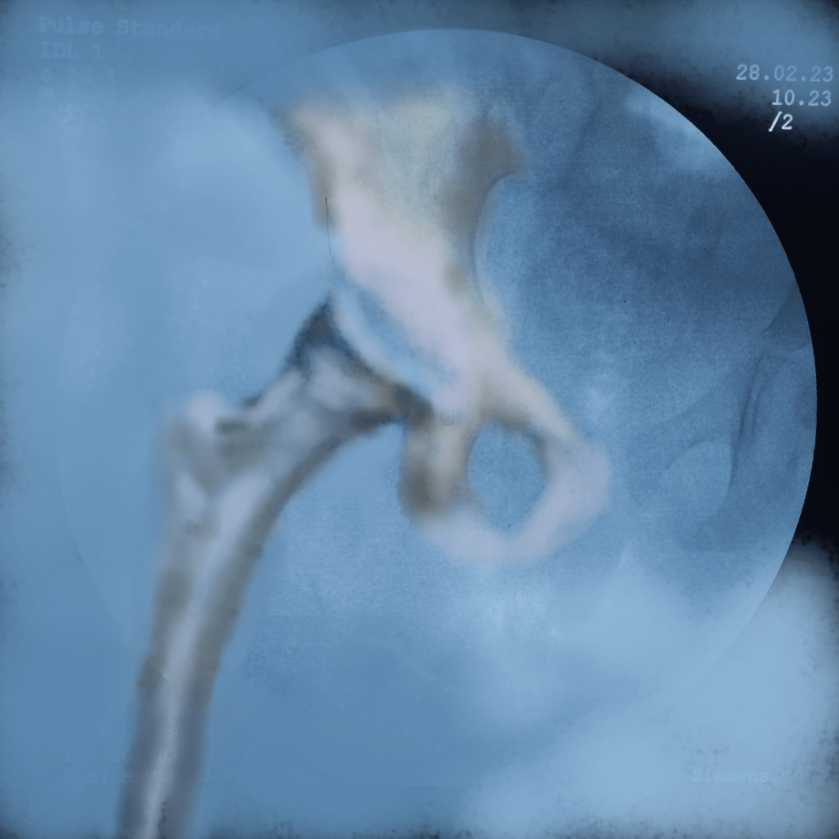
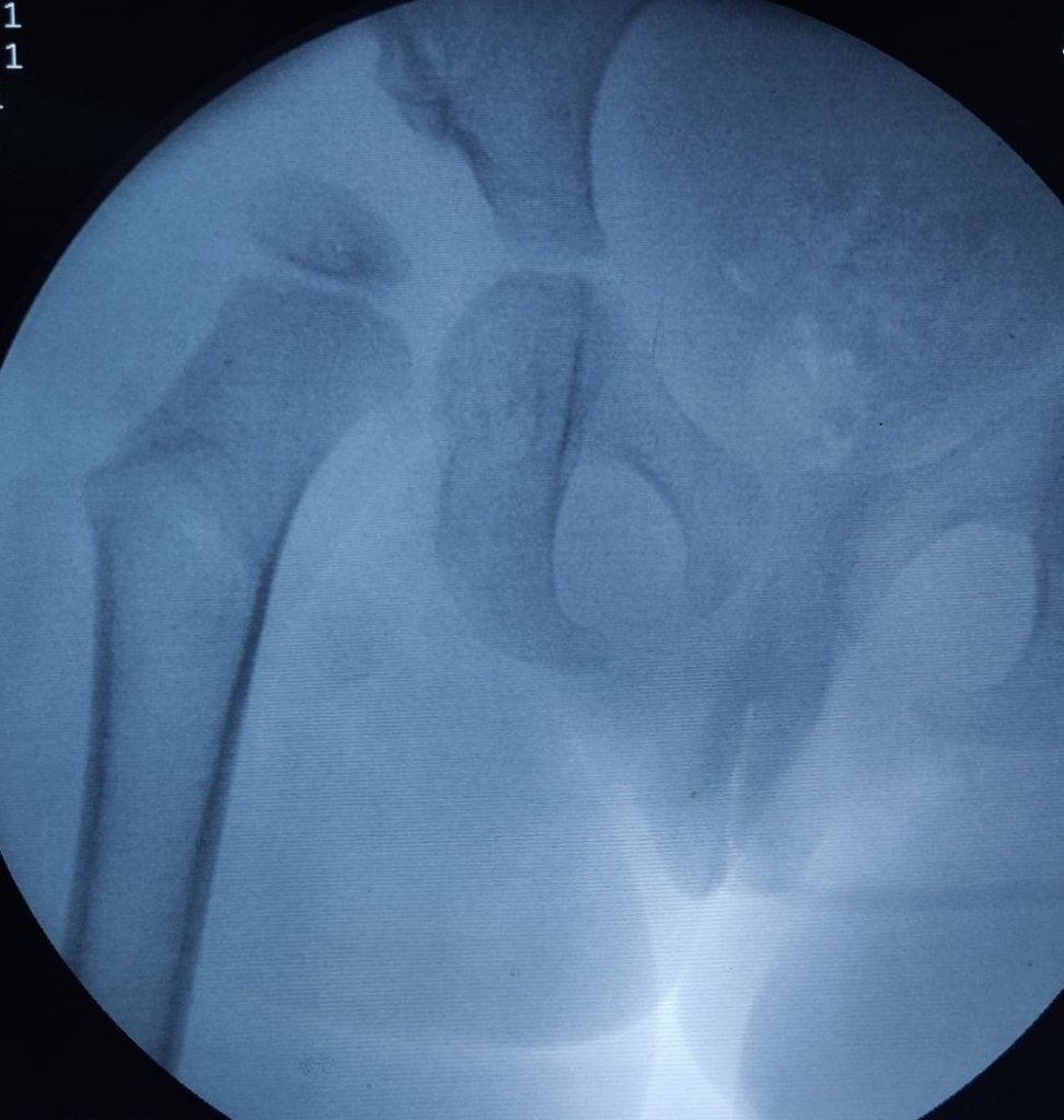
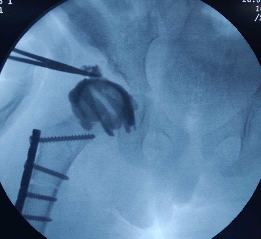
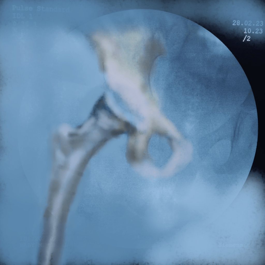
Arthrogram is an excellent tool to evaluate the state of cartilage over the lateral aspect and superolateral aspect of femoral head and then decide whether we want to do the acetabular osteotomy at the same sitting or defer it.
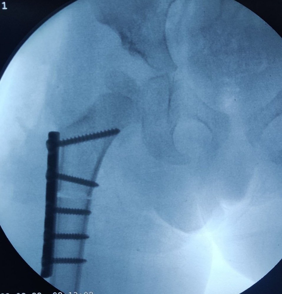
Illustrating below a case whether the Derotation osteotomy was done first and the we decided the acetabular osteotomy based on the cartilage cover on arthrogram.
This is useful in a child younger than 2 and half years as acetabulum has good remodelling potential at that age. In older children we will need to combine the procedures.
The state of hip before the osteotomy. Open reduction was done elsewhere about a year ago. Persistent hip dysplasia was observed. The Derotation osteotomy improved the coverage but we still have to decide about the acetabular procedure. An arthrogram reveals a large cartilage cover on superolateral acetabulum. The cartilage bump is pointed to by the forceps. The C Arm image is superimposed and an artist’s impression is drawn showing what the cartilage would look like in 3D. This offers an excellent teaching tool and a 3D orientation for young surgeons and pediatric orthopedic trainees to decide whether an acetabular osteotomy is needed
The surgeon would then discuss with parents and opt to continue an acetabular procedure of take a staged approach
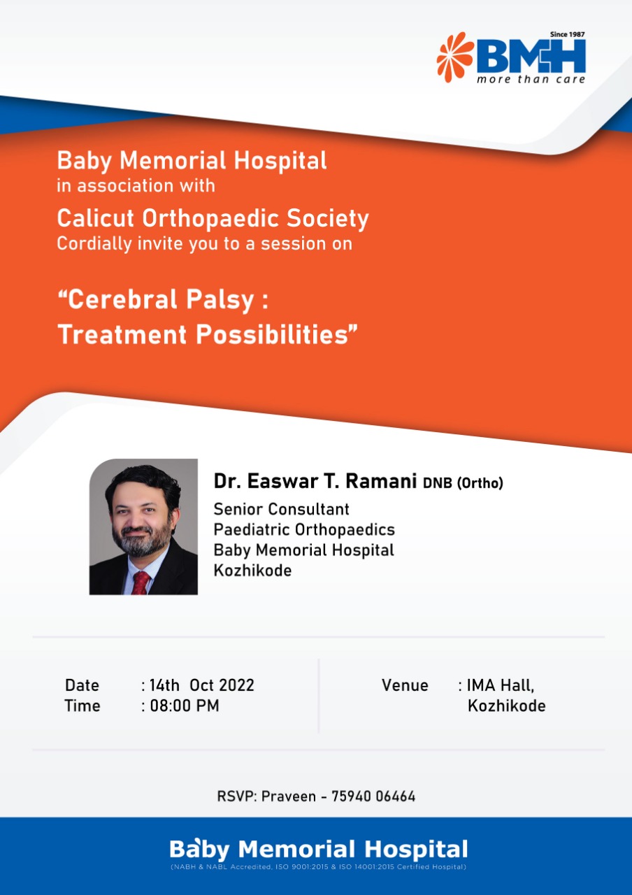
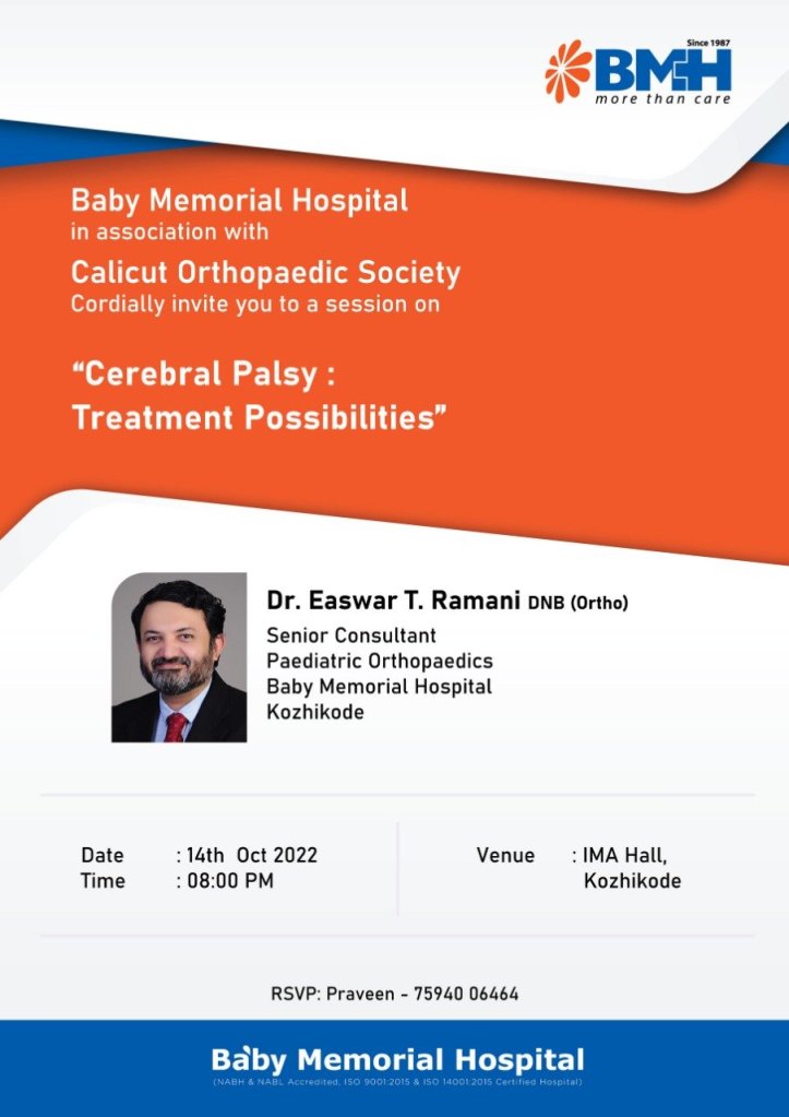
Excited to address the #Calicut #Orthopaedic Club this Friday and speak to stalwarts, teachers and trainees on #CerebralPalsy and the #orthopaedic treatment possibilities.
I will speak on the need for early detection, treatment and also about the newly released hip surveillance guidelines.
The trainees and general orthopaedic surgeons need to understand the importance of early referral and the benefits of early treatment.
In India with its exploding healthcare facilities and young population, we need to be aware, equipped and future ready when it comes to cerebral Palsy care.
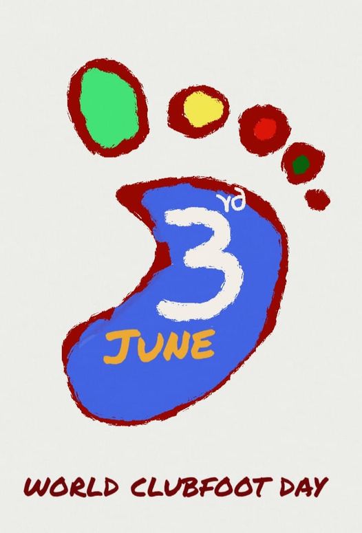
In years previous to the #COVID pandemic we at Palakkad Cooperative Hospital used this day and weeks around it to raise awareness, plan fundraising via MITRA Trust and re-dedicate ourselves tot he cause of Clubfoot care. Unfortunately there has been a hiatus in our activities for over 2 years.
Clubfoot Care Team at Palakkad District Cooperative Hospital, Palakkad, Kerala, India
This year we are re-starting everything.
What is Clubfoot ?
#Clubfoot is a #congenital#deformity of the foot which can be fully treated in most cases. The children lead a normal life after a successful treatment course.
Events
We will be oraganising events this year after a hiatus of two years to mark this day and re-dedicate our team at #Cooperative#Hospital#Palakkad to cost effective, accessible, state of the art and evidence based #Clubfoot#Care .
Medical Camp for Pediatric Neurology and Pediatric Orthopaedic Disorders
We are restarting our yearly free combined #PediatricNeurology and #PediatricOrthopaedic #MedicalCamp for #CerebralPalsy, #autism, #orthopedic #Diseases in #children this May at #Palakkad, #Kerala.
The children can consult a Pediatric Neurologist Dr. Velmurugan and Pediatric Orthopaedic Surgeon Dr. Easwar TR and also get counselling from Psychologist Mr. Toji Joseph and Physiotherapy consult from Mr. Biju Bhasker
The Medical camp is being organised as usual by the wonderful people at #Sevabharathi , #palakkad and also #LifeCarePhysiotherapyClinic, Palakkad Registration is free : 9495888879, 6238256073, 8891294916
Do share widely so that the news reaches the beneficiaries. Thank you Dr. Easwar TR Pediatric Orthopaedic Surgeon
Medical Camp for Pediatric Neurology and Pediatric Orthopaedic Disorders
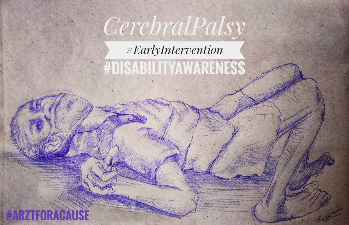
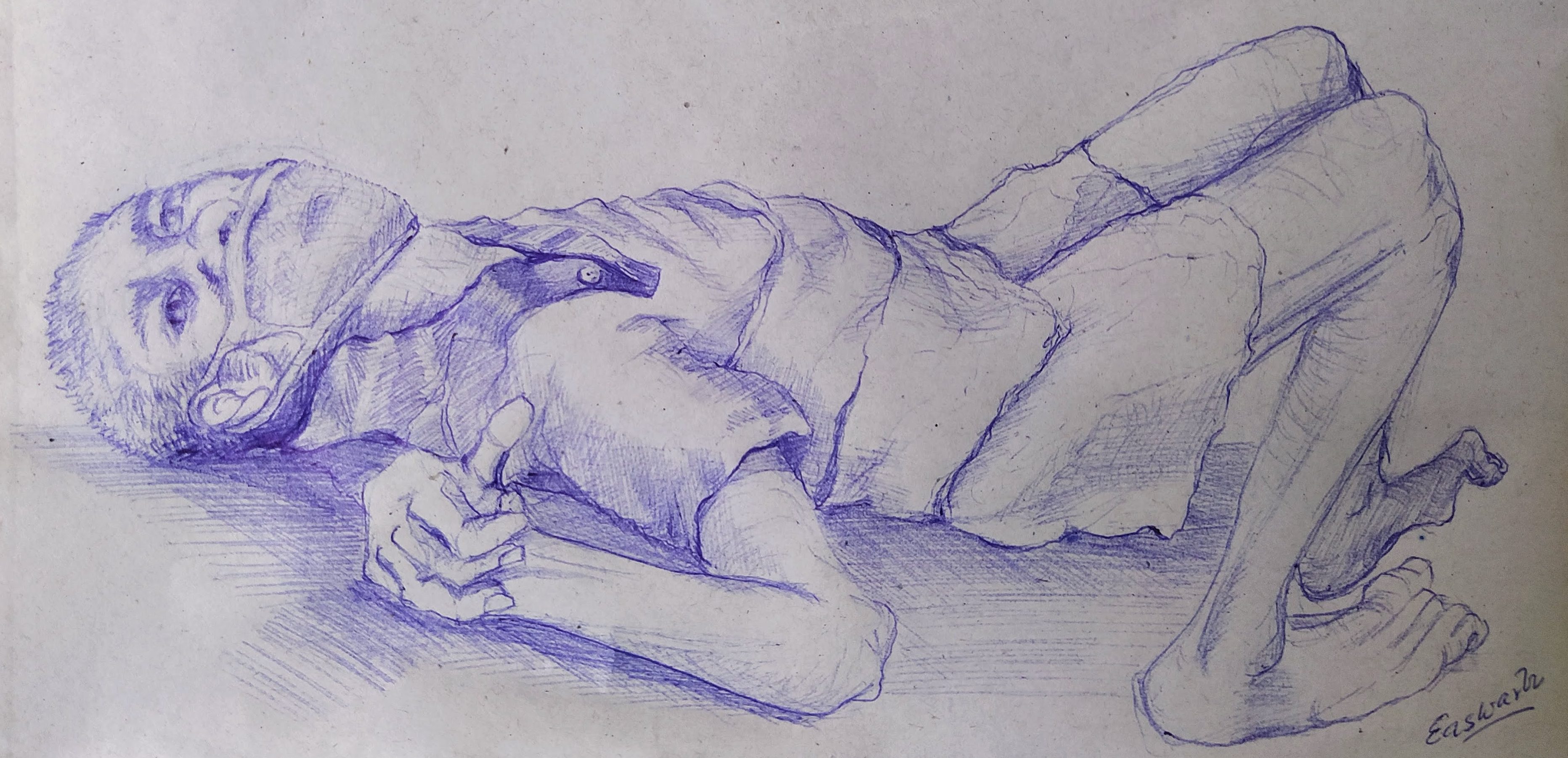
This is my #NFT #Art to raise awareness on #Disability caused by #CerebralPalsy .
Importance of detecting it early and early intervention is stressed. Quite often we find that children are not referred early and treated. This results in very bad deformities that are difficult to mend.
This is part of the #ArztForACause effort by doctors to improve disease awareness among public via 🎨#Art #paintings #nftart https://t.co/OzZUu9FJCh
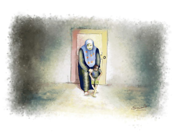
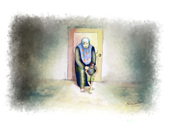
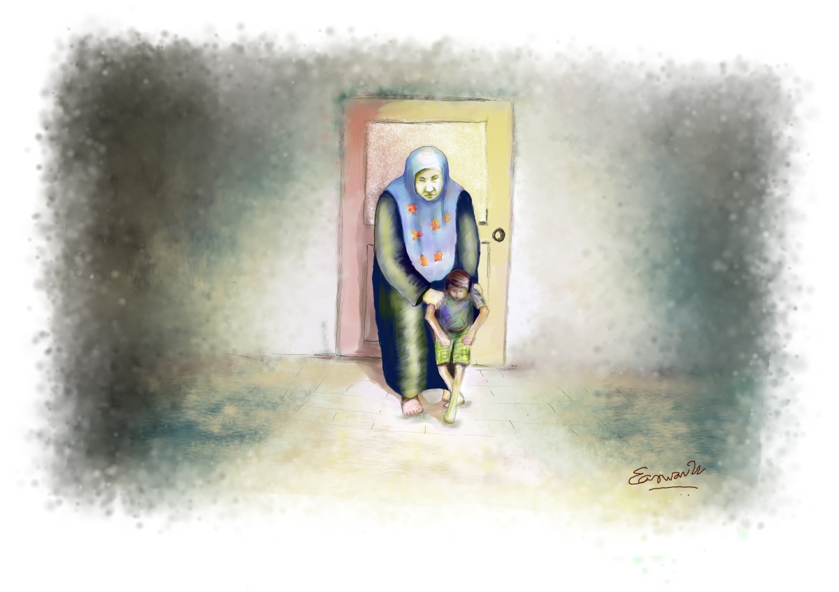

Cerebral Palsy is such a drastic turn of fortune for any family. The poorer the family, the more challenging. With her back to the closed door, symbolising all the lost life oppurtunities, a mother steadfastly leads her child along the well lit path of medical care with darkness of either side.
This tale repeats with so many children I see in my clinic. It is mind wrenching. Promise of quick cure lures any number of parents from that narrow path to dark streets of quackery and extorsion rackets who promise magical cures.
This is my tribute to so many parents who lead their child along the narrow lit path
Mother Leading a Cerebral Palsy Child
Even when treatment of cerebral palsy is widely available, it is not uncommon to see severe deformities due to improper or delayed referral to a treating centre, lack of awareness of simply neglect. Quite often patients live in far flung villages with poor access to healthcare or broken families compounding the difficulties.
Advanced deformity of the hand in Cerebral Palsy
I have seen that through these storms invariably the mother stands and guides the child unwavering.
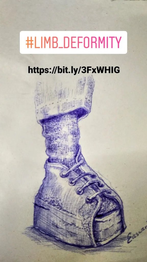
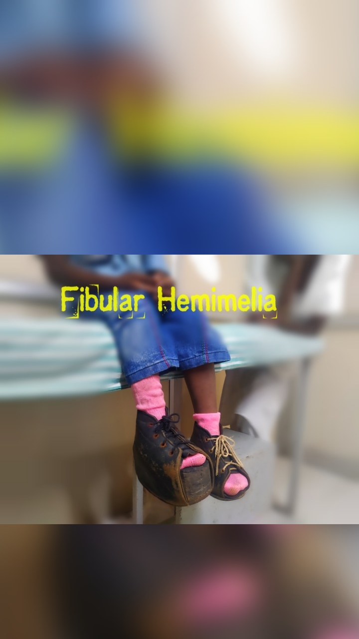
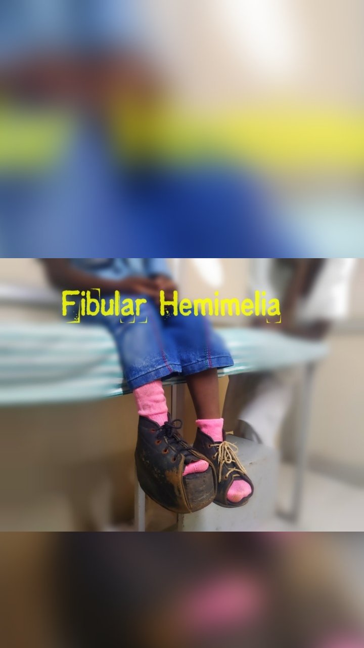
#FibularHemimelia #NFT #Art for #WorldDisabilityDay
As part of our #ArztForACause effort to raise awareness of #PediatricOrthopaedic #Diseases and upcoming #WorldDisabilityDay I have made this #Art on #FibularHemimelia
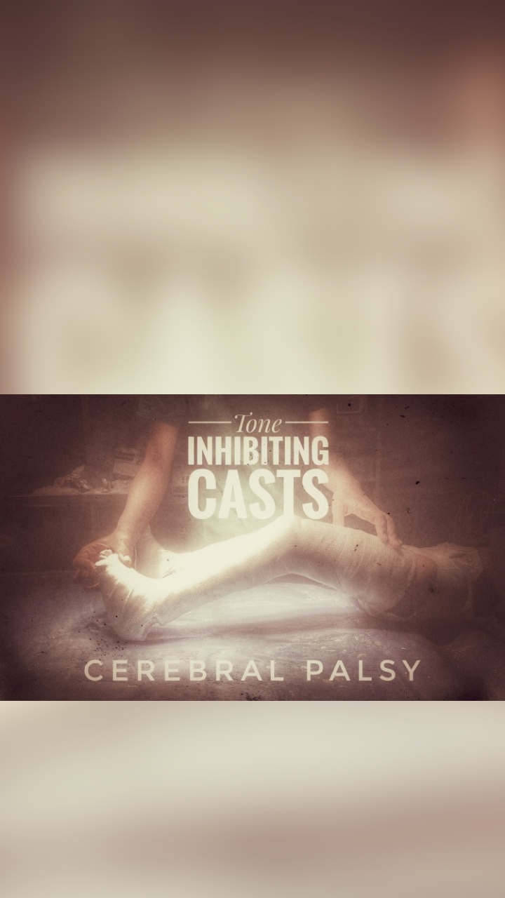
Cerebral Palsy is a tough problem in Paediatric Orthopaedics. The children have spasm, contracture and variety of coordination issues , balancing issues, seizures and other symptoms like tremor, dystonia which makes treatment quite a challenge. This is more so in the younger children where we would like to have a tool that is non invasive and still helps relieve spasm.
Tone Inhibiting casts offer an invaluable tool to control spasm in a very young child that you would want to avoid surgery.
The dosage of surgery in cerebral palsy has to carefully titrated as muscle are inherently weak and over-lengthening can be disastrous.
We try to avoid overzealous initial surgery and but time when possible with plaster casting to reduce muscle spasm.
Tone Inhibiting Casts are a vanishing tool these days in medical professionals’ armamentarium but find it a great tool. In India with cost of Botox injections to relieve being very high and in the class of patients I treat many of who are from poorest strata this is a godsend.
Cast application is done under general anaesthesia so that adequate muscle relaxation is obtained and we take great care for a close , bespoke type fit with good pressure relief over bony prominences. Prevention of pressure sores is very important and technique of cast application – the fit, padding, tightness, joint position, strength of cast all play a role in the final result.
We keep child on physiotherapy all through the time child is on cast to keep up muscle strength and also aid stretching. A cast usually is kept for 6 weeks. Sometimes in severe spasm we have staged application of casts to progressively apply it lesser flexion of joints as the muscle tone decreases.
Back Pain Clinic - Adults and Children, Palakkad, Kerala
Happy to announce that the Spine Clinic for Children and Adults will be functioning on all days except Thursday at Palakkad District Cooperative Hospital & Research Centre, Palakkad, Kerala.
This will cater to all variety of Spine diseases in Children and Adults.
Common diseases like Disc Problems, Infections of Spine ( Septic Spondylitis ) , Tuberculous Spondylitis, Scoliosis, Spina Bifida are treated here.
I am available for consultation on all days except Thursday from 10am to 4:30 pm. Please contact Palakkad District Cooperative Hospital for details and appointment.
My Clinic restart has been delayed due to Covid situation and we have noted that children with fractures are having difficulty getting consult for emergencies.
A speciality Fracture clinic for Children run by Dr. Easwar TR is operating at Malabar Hospital Palakkad.
Treatment is provided for all types of children’s bone problems, sports injuries and fractures. The service is available around the clock in association with Orthopedic doctors at Malabar Hospital, Palakkad.
For Emergencies and Appointments please contact Malabar Hospital for appointments.
Children’s fracture clinic for bone and joint problems, fractures, sports injuries at Malabar Hospital Palakkad Kerala by Children’s Bone Specialist Dr Easwar TR
You will need a free account to get started. Registering is simple and free. Get yours at https://medtatva.com/signup. Remember to read the Terms and Conditions, Privacy and Disclaimers before using the App and the website. Kindly keep in mind that Privacy policy when uploading data / personally identifiable X-ray on the website.
We are all going through a tough time with #COVID19 #Epidemic. It has been a testing time for doctors, public and administrators alike. The poor have been hit badly and are under threat as the epidemic is still spreading in various countries as we speak. Access to healthcare, paying capacity, travel are all issues that the citizens of various demographics have to confront.
We at MedTatva [ https://medtatva.com ], have been trying to solve accessibility to healthcare and diagnostic tools by using technology.
Past few days we have put together the best recommendations from CDC and WHO to build a symptom checker that is simple and which could be completed in less than a minute by a non-medical person.
Cerebral Palsy is such a difficult disease to handle and treat for the health care professionals.
The treatment process needs to take the parents and family members along so that these children get optimal care. It’s important to reinforce on the parents and lift their morale throughout the course of long and arduous treatment.
I routinely arrange seminars and medical camps and arrange public events in association with groups serving these children to help them and keep caregiver morale high. It’s a fight, a relentless fight against Cerebral Palsy.
It never ceases to amaze and move me that the mother , however humble or rich , illiterate or well educated, they may be cares for the child and stays with child through thick and thin.
Wacom One CTL and MyPaint on Linux works like it’s a match made in heaven.
My Digital Art on a mother caring for her child with Cerebral Palsy – Drawn using Wacom One CTL 472 and MyPaint Open Source Software.
This painting is part of the #ArztForACause fundraiser for children with Cerebral Palsy , Autism and Movement Disorders
She literally is the guiding light of the treatment, showing the way and proving that she’s the true representative of the all giving , ever loving Divine Mother
A Mother leading and lighting the way of care for her child with Cerebral Palsy – Digital #Art
World Cerebral Palsy Day - at Palakkad and Shoranur, 2018
#WorldCerebralPalsyDay is afoot. Like last year, I am planning to conduct month long events across Palakkad and Shoranur to raise parental awareness on treatment of #CerebralPalsy.
Most of the parents who come for care of their children come late, unfortunately with joint contractures, hip subluxations, alteration in knee mechanics and foot deformities. Aside from these the problems these children have with upper limbs, hands and doing activities.
Many of these children have uncontrolled seizures, terrible difficulties communicating, speaking out their issues apart from ambulation. Ambulation may in fact be least of their issues. This sad fact goes to show how important early referral is and how important it is to enroll the child under a care of a Pediatric Neurologist, Pediatric Physiotherapist who can then co-ordinate care with their colleagues in Pediatric Orthopedics, Pediatric Medicine, Occupational Therapy, Speech Pathology, Audiology etc ..
Cerebral Palsy care is a great team effort. No part of the team is greater than other, but the role of the General Practitioner / Pediatric Medicine Doctor, Pediatric Physiotherapist is central, at least in India. They determine the sole referral entity, a person with whom the parents spend their maximum time and trust with. They are the ones in whom they confide most of their issues with.
World Cerebral Palsy Day is a wonderful concept to reinforce these simple care giving principles. Parent education and empowerment being central to the cause of care of these children. Like 2017, we are organising activities across the district of Palakkad – in Palakkad town and neaby rural areas and Shoranur town to educate parents and also conduct early detection and intervention camps to identify children who need care.
World Cerebral Palsy Day – at Palakkad and Shoranur, 2018
Activities planned :
Free medical camps for underpriviliged children
Special clinics with Gait Analysis
Funded Orthotic programme for poor children
Pediatric Physiotherapy advice
Custom made Wheel Chair fitting
Parent awareness of Cerebral Palsy – lectures and symposias in rural areas
Notices and public handouts to improve general awareness
Involvement of other NGOs , social service organisations to improve public outreach
These activities would be done throughout Ocotober 2018 under aegis of the Cerebral Palsy Clinic at Rajiv Gandhi Cooperative Hospital & Research Centre and ICCONS, Shoranur
Contact Numbers for Clinic bookings and more information:
As a practicing Paediatric Orthopaedic Surgeon, I am called to meeting to present my work. This involves presenting to peers, co-workers, patients and parents of children I care for. Each of these presentations will be with a different focus on a particular topic.
While this is not an uncommon scenario, the solution to create a reusable presentation slides using #OpenSourceSoftware tailored to individual audience is. Most doctors are not familiar with programming environment and shy away from anything that is not WYSIWYG. They rely on good old #PowerPoint / #Keynote to save them. At the most some of them may try and use the clunkly #LibreOffice or #OpenOffice if they want to stick to OpenSource. Recently with advent of tools like Prezi, media heavy interactive presentations have become popular. The popular presentation softwares of KeyNote, PowerPoint have also spruced up their animations and transitions to enable them to look more attractive. Still the WYSIWYG nature of these and point-and-click makes them very slow. We could achieve better, faster and more attractive results with using #FOSS tools.
FOSS Image and Video editing softwares as required to arrange the media. I mainly use GIMP, InkScape, KdenLive, OpenShot, HandBrake to arrange my media and encode them.
My choice depends on the demands of the presentation.
For media heavy, especially video heavy presentations I use RevealJS. For presentations that are more of less static with few videos I tend to use Beamer / LaTeX. What I note below are my experiences as I tried to create a smooth workflow that could replace PowerPoint ( or KeyNote / LibreOffice ) as a tool to create #Medical Presentations.
Overtime I have refined my workflow and now I find that I am far more productive and my slides can pack much more information than a power point slide. While the more advanced interactions would require some knowledge of JavaScript, and therefore would turn off most doctors, most of what follows require minimal programming use.
I will detail my workflow to create simple fast layout using Emacs and RevealJS without handcoding of JS and HTML. We will be relying on the RevealJS, Emacs, Org-Mode and ox-reveal package to do the lifting.
Disclaimer : Even though it doesn’t need programming knowledge, ability to use Emacs is a must for this workflow. It is preferable that one is on a Linux OS as the attempt is to go all #FOSS here.
so, here goes ….
Aim
To create an visually impressive medical presentation using non Power Point open source (FOSS) softwares.
Tools
Emacs (24.3 or greater)
org-mode
org-reveal
Reveal.js
Chrome Browser
Open source video codecs on the system
Why this and not PowerPoint ?
Over the years Medical Conference presentations have got mature and old tools have got boring. Varied audiences, topics, media content , interactivity required, transitions and animations to keep audiences interested have all changed.
PowerPoint with it’s traditional set of tools is boring to say the least. The point and click interface is slow by comparison to plain text typing. This seems counter intuitive to PowerPoint pandits but I’ve found that once the media is arranged and readied, once can create more far more attractive presentations with the tools mentioned above.
As far as medical presentations go, the video presentations embedded PowerPoint / LibreOffice have a habit of breaking on stage. I have seen numerous instances of this happening.
And, of course PowerPoint costs 💰💰
It is also Closed Source making it difficult to edit and reproduce when you are with a system where it is not installed.
Okay, but why Emacs, why indeed ?
Emacs is Open Source
Emacs is stable
Emacs is good
Emacs is better than #Vim
Emacs has un-paralleled number of extensions and programming support
Emacs has Org-mode…
Okay, So why org-mode, what has that got to do with presentations ?
org-mode is cool
org-mode is simple text
org-mode can be manipulated anywhere with text-editor
Its FOSS
It can be extended with other tools like org-reveal
Hm, Okay, but why Reveal.js ?
An actively developed FOSS Tool with a community
Allows 2D stacking of slides permitting nesting
Plugins and all the JS/CSS/HTML5 goodies can be integrated
Very good slider-presenter notes
PDF export option for handouts
Very nice transitions and animations
Good builtin themes and literally infinite customisation options as per CSS
Works very well with slide-projectors and remote tools to advance slides
Okay, but why use org-mode / org-reveal with Reveal.js ?
Org-mode is cool, easy, transparent text typing
org-mode is structured and nested just like a regular presentations would be
One can easily do a text-only sketch of a presentations by typing out a few lines of text in org–mode formatting and out put a neatly animated stacked presentation in Reveal.js
If one were to code HTML and JS with Reveal.js, it would be considerably opaque, with HTML markup and JS obscuring the structural details of the presentation.
By integrating org-mode, org-reveal and reveal.js we are integrating all that is good in respective tools while sticking to what the non-programmer user ( an average medical professional ) would like to do – type text and structure the presentation.
Note down the path to the folder relative to the folder where the presentation will live.
If you have Bower installed you can just do bower install revealjs
Step 6 :
Create a folder where your presentation will live.
Inside the folder create subfolders for Images, Videos, Scripts, CSS styles and other documents which may be needed for the presentation.
Now we can create the main file of the presentation – the Org-mode file using Emacs. Org-mode file is a simple text file which can be opened using any other text-editor. It has the extension of “.org”.
While using Emacs and Org-Mode, however, it provides lot of goodies. Org-Mode in Emacs has lot of extensions one can install that extends it functionality. One can for example use the same org-mode file to output HTML, LaTeX, and PDF.
So let us create the main presentation file. I title my presentations the following way, giving it context, separated by underscores : <topic>_<audience>_<date>_<venue> . For example if I am giving a public talk on Cerebral Palsy at my home town of Palakkad, on July 30th,2019 , I would title my presentation like this : “CerebralPalsy_PublicTalk_Palakkad_30072019.org”
This allows me to keep separate org-mode files for different audience and keep using the same images, videos etc.. Therefore I am fully portable and self-contained when I have to whip up a presentation tailored to any particular audience – technical or non-technical.
C-x C-f in Emacs to the file you want to create with .org extension.
C-C C-# to insert Template for a Reveal.JS presentation.
If you have ‘ox-reveal loaded it should be available as a choice.
Once chosen it will list some options at the top of the org-mode file.
We will need to provide the path to the REVEAL_ROOT directory to the place we have stored the reveal.js library. This path is relative to the folder where the file for presentation lives.
Once these are done, It is important to get the images, videos ready. They have to be edited using FOSS tools for editing photos and videos. Once edited they’ve to be named properly so that we can reference them in our presentations.
This completes the ground work required to start writing the presentation. While this may seem a lot of work, one must remember this is one time effort.
We will deal with the actual creation of org-mode file, the options while using Reveal.JS in the next part ….
Developmental Dysplasia of Hip - Left hip is dislocated
DDH otherwise called Developmental Dysplasia of Hip is a condition where the ‘ball-and-socket-joint’ of the hip is not formed well at birth. It used to be called Congenital Dislocation of Hip.
The child is born with a slightly misfitting hip joint where the ball is slightly shifted out of cup or a hip joint where the ball is completely outside the cup and cup is also very shallow. This can occur on one side or both sides.
Developmental Dysplasia of Hip – Left hip is dislocated
Why does it happen ?
Most times its ‘Idiopathic’ , a medical term which means – cause is not known. In many other patients, the child will be having one of the syndromes (a group of clinical features and disorders in other organs systems and external appearance) or neurological issues in the spine (like meningomyelocele) that also causes this. This second group is sometimes medically referred to as Teratogenic DDH and is much more difficult to treat.
We will discuss below the aspects as related to Idiopathic DDH. They don’t apply to Teratogenic or Neurological DDH.
How do we know the child has DDH ?
Most newborn screening procedures at the hospitals have doctors checking to see if the new born child has clinical signs of DDH. It is easiest to detect and treat it at that early stage.
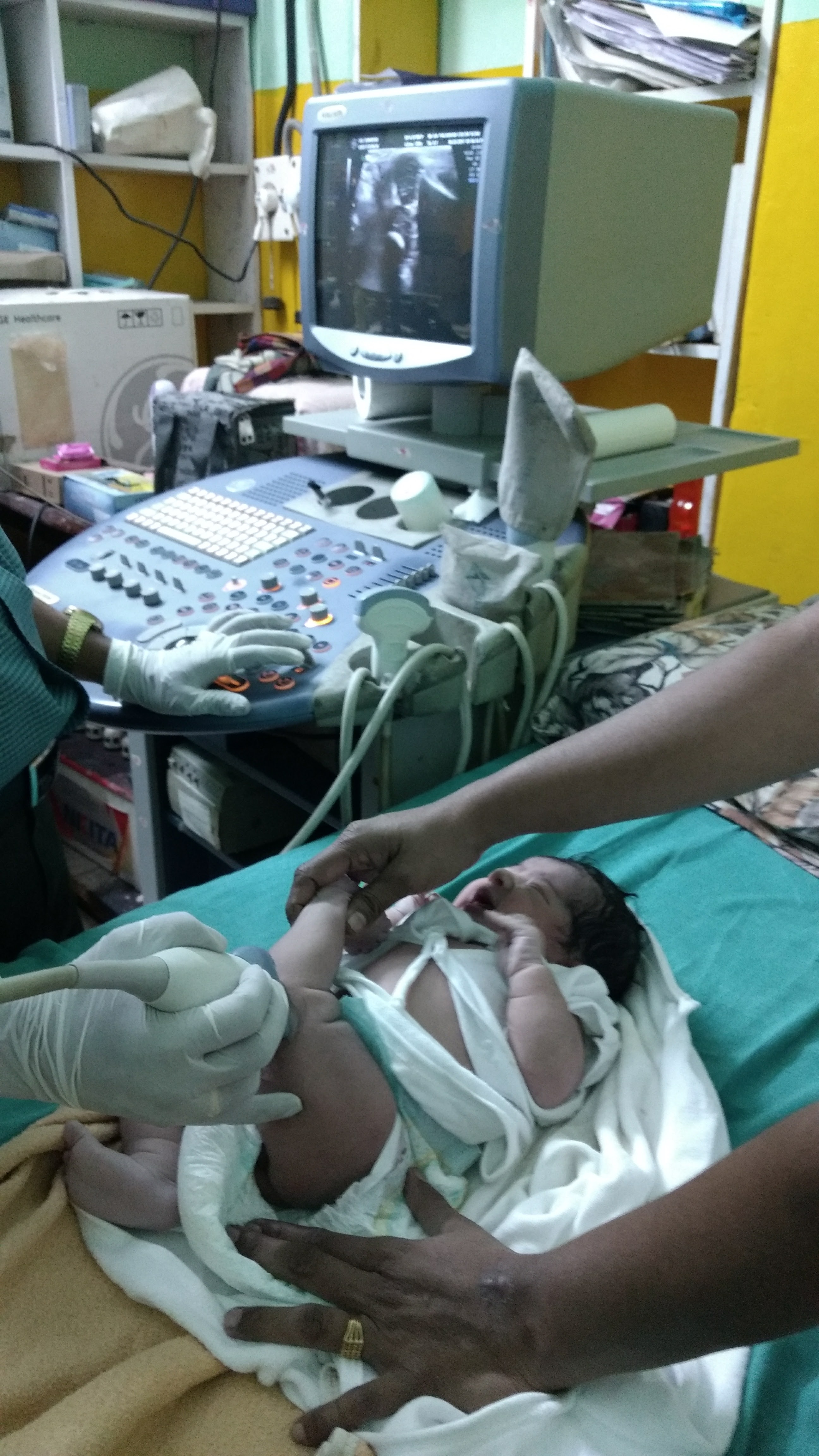
Once a clinical suspicion of DDH is there the Neonatologist usually refers the child to a Pediatric Orthopedic Surgeon. In addition to establishing the clinical diagnosis by physical examination, the doctor will order an x-ray and an Ultrasound Scan of the Hips to check the dislocated hips. Several measurements need to be taken and ease of dislocation need to be established under Ultrasound scan by a procedure called Dynamic -Ultrasound. The treatment starts after these are done.
In many children, the initial diagnosis may not be made in the immediate post-delivery period. Children may be brought to the OPD by parents with complaints that the thigh skin fold dont look symmetrical and the hips don’t open out fully for parents to change diapers.
In older children parents may notice a limp when the child walks or a limb length difference may also be noted with the dislocated side being shorter.
Limb Lengths may be noted to be different in DDH when child is older and walking. The limb with the dislocated hip will appear shorter.
How soon should the treatment start ?
As described in previous section, treatment should ideally start in neonatal period once the problem has been detected. After clinical examination and Ultrasound examination, your doctor will suggest usually a belt type device called Pavlik Harness to be applied on the child to keep the hip reduced inside the cup. This harness is to be worn full time. Parents are counselled on how this is applied and how the straps are tightened.
Ultrasound exam for a child with Hip and Knee dislocation
Once this harness has been applied, the hips are scanned with Ultrasound every 2 weeks to check for improvements. Ideally this has to be kept for about 3 months and taken off after the hip stabilizes inside the socket. A slow weaning period is there before the child can be fully taken off the Pavlik Harness. This is to ensure that the Ball ( head of femur ) stabilizes inside the socket (Acetabulum of Pelvis) and the hip is stable. The tissues around the hip also has to shrink and adapt to the new location of Head of Femur. Otherwise re-dislocation or partial slip (medically called subluxation) is a possibility.
In many cases the parents may not be willing for a device to be applied on the child full time post-delivery or the DDH itself may go undetected. This child eventually may be brought later in life by the parents with complaints necessitating surgical procedures.
What is the treatment options for DDH?
As previously described if the newborn is detected to have DDH the treatment is to apply Pavlik harness on the child after Ultrasound exam proves hip is reducible and stable in what is called a safe zone. Safe Zone is a zone where the position in which the harness has to be applied to the child does not compromise the blood supply the head of femur. This is very important decision to make.
Pavlik Harness for DDH hip needs to be fitted properly
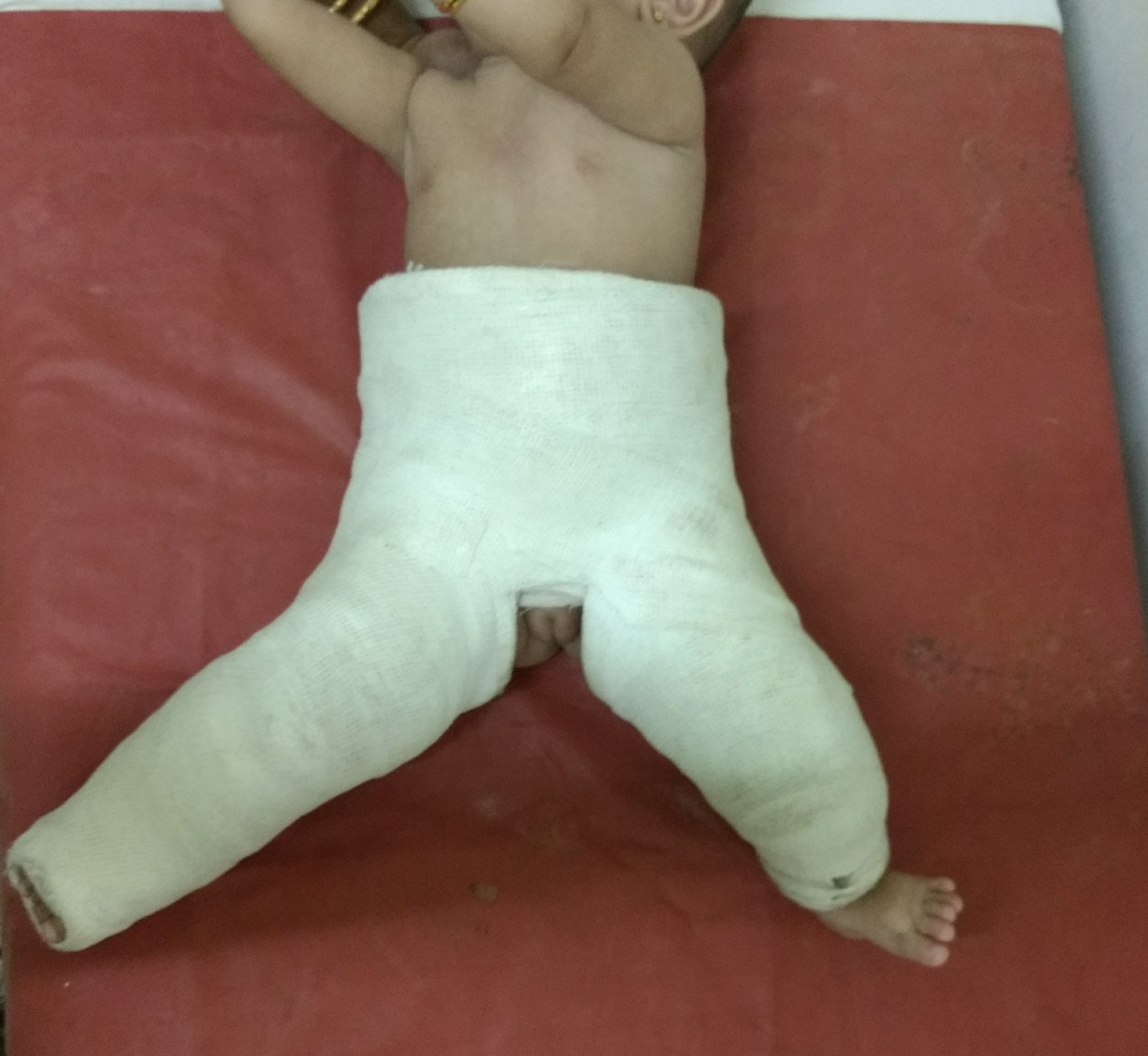
Hip Spica Plaster in child with DDH
Application of Hip Spica under anesthesia in DDH
If the child is brought later in life, before 1 year, the hip has to be checked for reducibility as the tissues around the hip would have become contracted and sometimes it is difficult to reduce the hip back into the socket. Usually anaesthesia would be required and the hip once reduced back into the socket is held there by application of the plaster of paris or POP called Hip Spica. This is kept for approximately 3 months until the hip has stabilised inside. A CT scan to verify whether the hip is reduced should be done while child is in POP as slippage of the hip socket is also seen when POP cast becomes loose with time.
Surgery is the only option in older children with DDH. These range from relatively simple procedures to complex hip reconstruction procedures depending on age of the child, slope of cup, angle of rotation of the neck of the thigh bone and shape of the head. These are best evaluated as per the needs of the child.
Arthrogram for DDH
Generally these surgical procedures can be grouped into a few types :
Open Reduction of Hip and Capsulorraphy (medical term meaning stitching up of capsuleor the covering around the joint)
This is basic necessity. Here the hip is reduced back into the socket and the covering, the capsule, which is loose is tightened with several sutures
Femur Derotation Osteotomy with shortening
This would involve a slight shortening of the hip bone and turning it around. The bone would then be stabilied with a metal plate and screws
The need for your child to have this done depends on the age of the child and the angle of the ‘neck of the femur’, something medically known as Anteversion. This procedure essentially de-rotates the femur neck and allows the head of femur (the Ball) to deeply sit inside the Acetabulum (the socket). A slight shortening of the thigh bone may be needed as the child , if old enough, would have developed tightness of the tissues and hip should be sitting inside the socket slightly loose than in tension.
Acetabular Osteotomy
In many children, when they present to the clinic their Acetabulum would be shallow and not deep enough to receive the Head of Femur. In such children deepening of the Acetabulum by a surgical procedure called Osteotomy (a type called Salter Osteotomy being the most popular) may be done.
The above procedures may be combined along with Open Reduction as the surgeon sees fit after evaulating the child. These options and its pros-and-cons would be discussed with the parents before the surgery.
What happens if we don’t treat DDH promptly ?
Best time to treat the child is in the newborn period. With time the tissues around the hip gets tighter and the bone structure of thigh bone and pelvis alters since the normal growth with moulding of the ball being inside the cup doesn’t happen.
This lack of moulding results in a shallow cup. Additionally the thigh bone’s upper end would be twisted out of shape and the ball also would be misshapen since it has not been moulded by the acetabulum.
All these result in a much more complex procedure to try and restore the hip.
Left untreated DDH causes osteoarthritis or the Hip joint and Hip pain in early adulthood often requiring complex reconstructive procedures.
It is best if the treatment is not postponed and done at the earliest.
Hip Dysplasia Awareness Month – June !!
The Hip Dysplasia Awareness Month of June is to educate parents on Hip Dysplasia and its treatment methods. We hope that parents would bring children to care centers at the earliest if suspicion of hip dysplasia is there.
A free medical camp for underpriviliged children suffering from Cerebral Palsy and Pediatric Orthopaedic disorders was organised by Dr. Easwar T.R and the administrative team at ICCONS, Shoranur, Kerala on 24th, May,2018.
A gamut of problems commonly seen in children with Cerebral Palsy was observed in OPD. While some parents had the good fortune of seeking medical care and intervention early in course of disease and others were not so fortunate. Lack of parental knowledge, distance / accessible care, financial cruch, normal siblings to care for and single parent challenges were the most important issues observed on casual data collection.
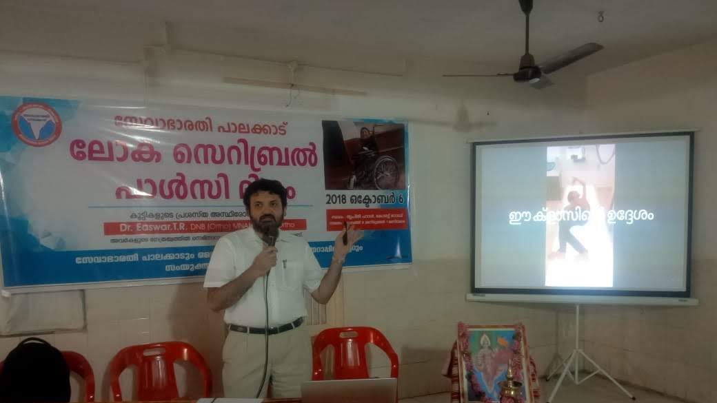
The medical camp was followed by an awareness class for parents, therapists and other care givers by Dr. Easwar T.R on the various aspects of Cerebral Palsy.
Custom wheel chair and mobility solutions for needy patients was discussed with Physiotherapists and low cost solutions for the same identified. Mobility solutions continue to be a major challenge for children in rural areas with poor roads, small homes with narrow doors and steps inside homes especially with outdoor toilets.
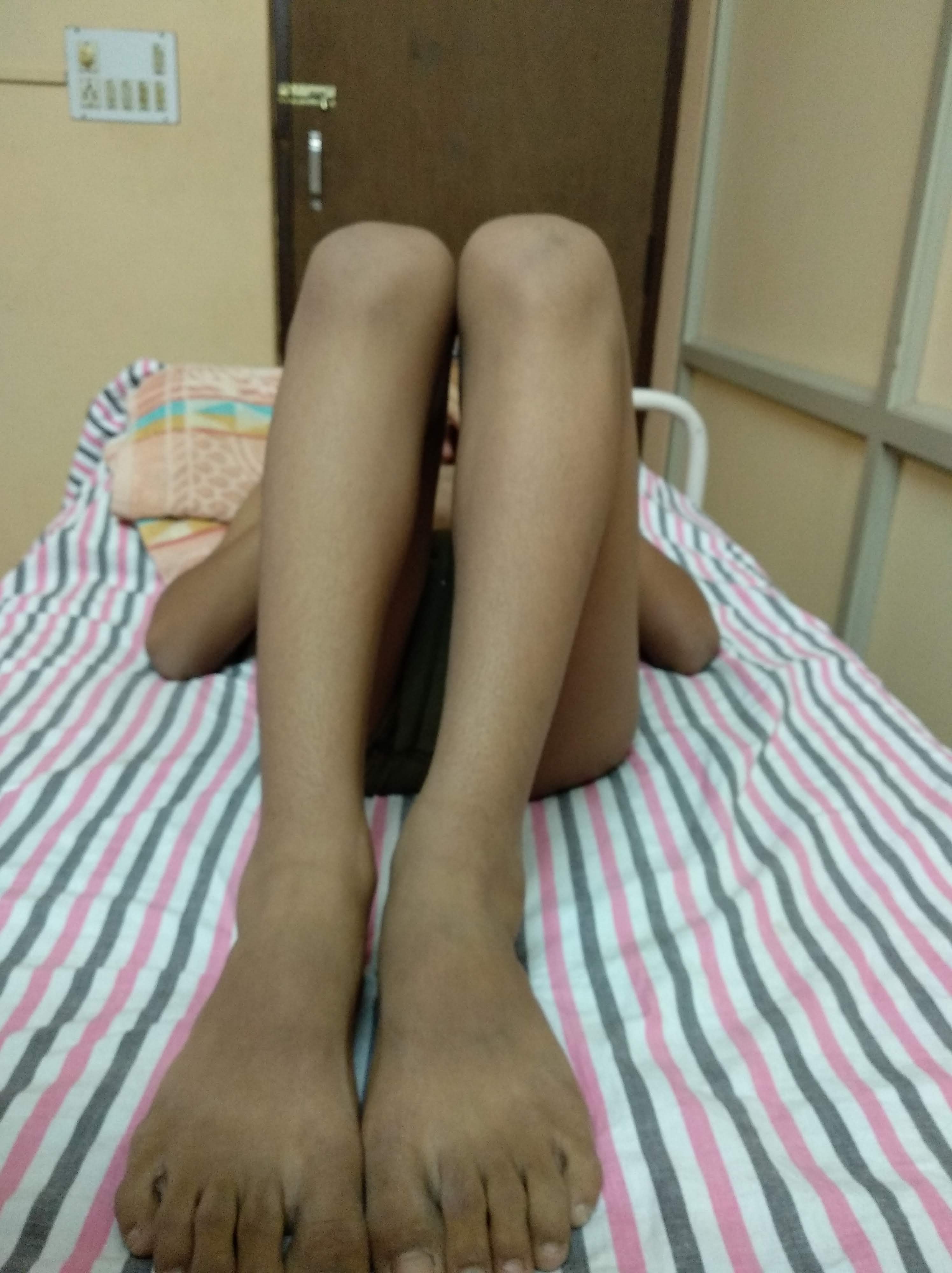
Flat foot in Cerebral Palsy
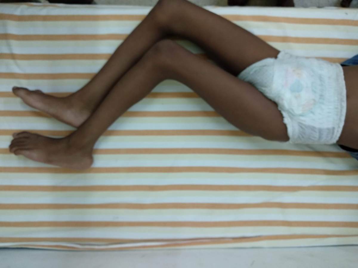
Cerebral Palsy child with widestance and flat foot
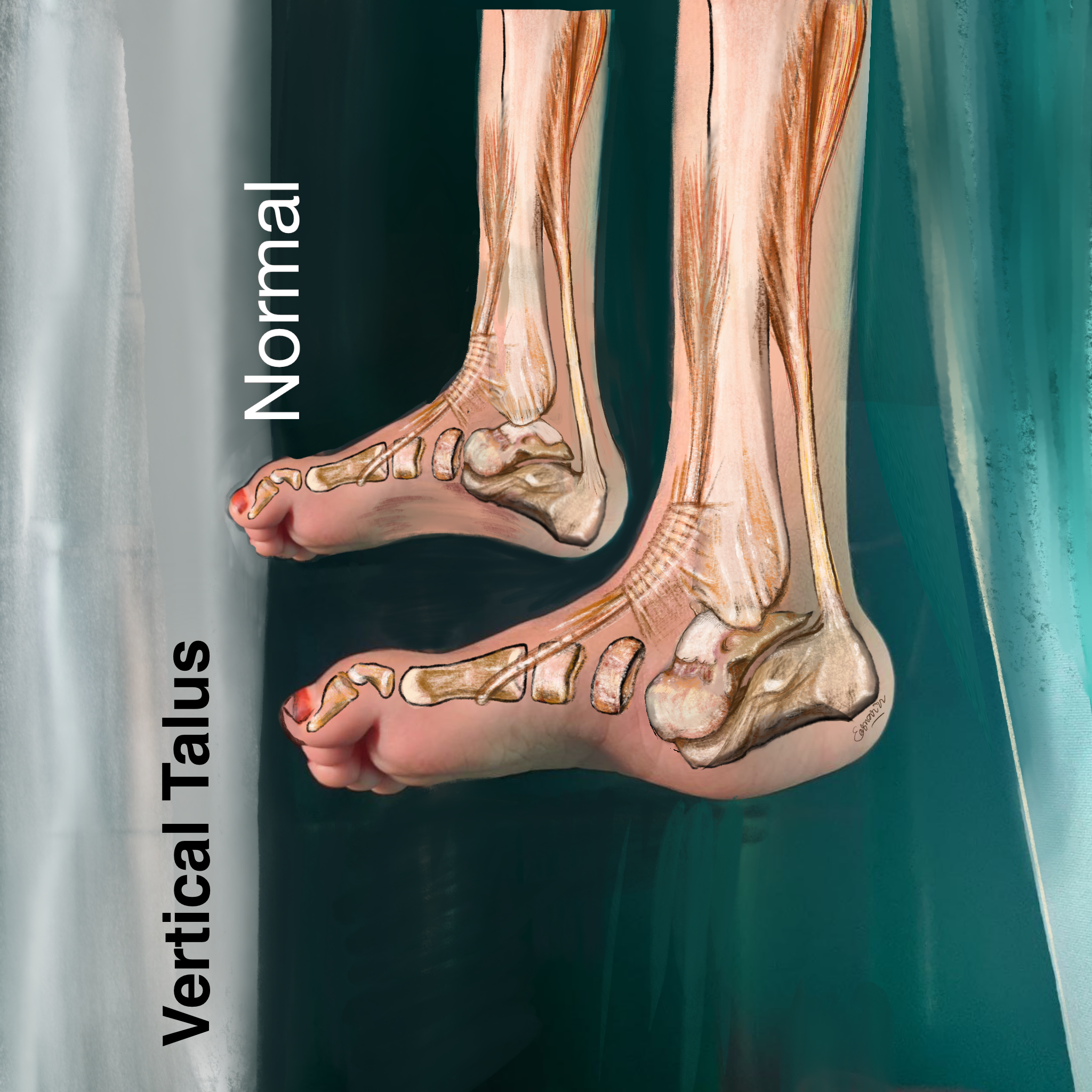
Hearing that your newborn has a foot deformity can be overwhelming for any parent. If your child has been diagnosed with Congenital Vertical Talus—often referred to as “rocker bottom foot”—it is completely normal to feel anxious about what it means for their future.
The most important thing to know is that this condition is highly treatable. With early and proper intervention, children with vertical talus go on to walk, run, and play just like their peers.
Art for Awareness: The ArztForACause Initiative
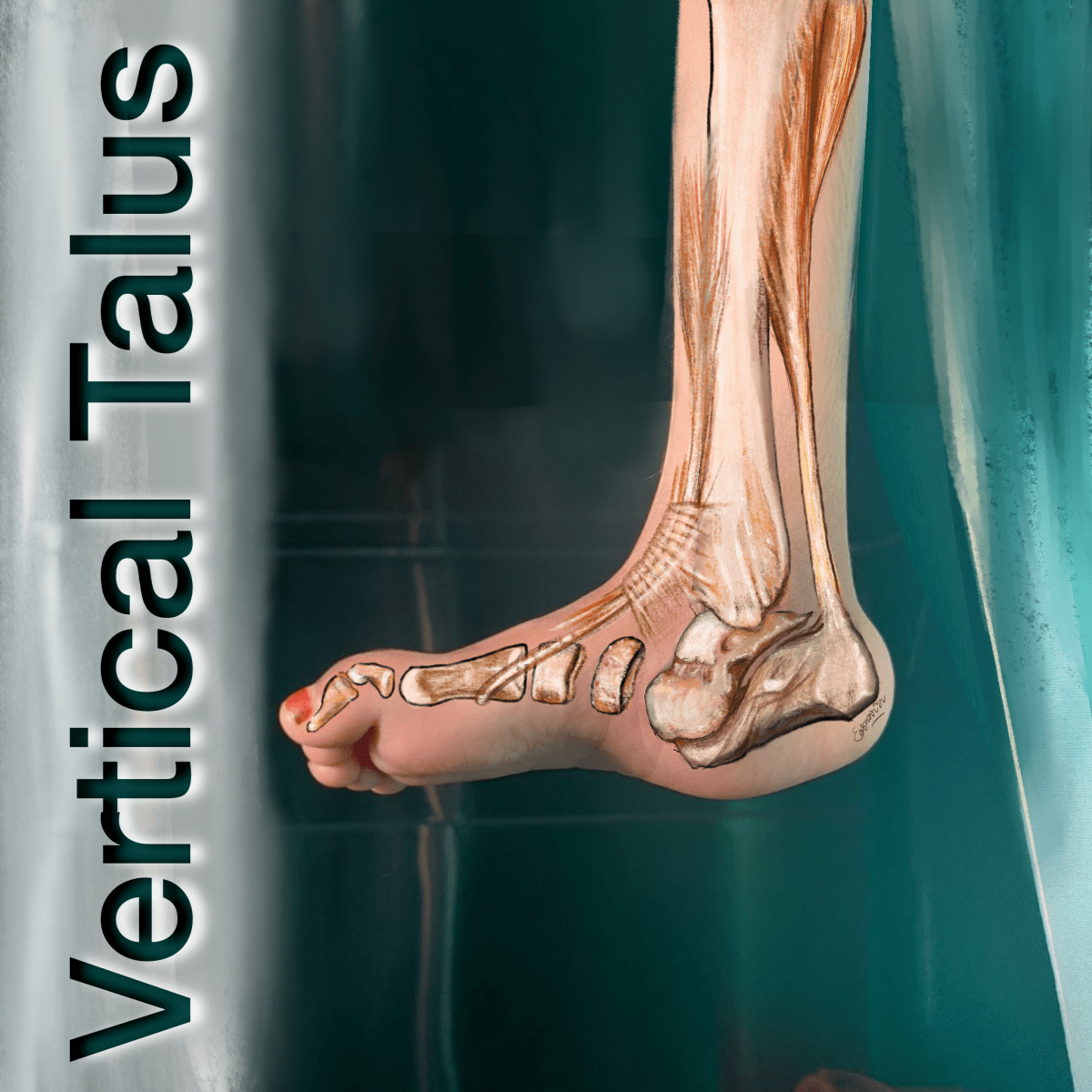
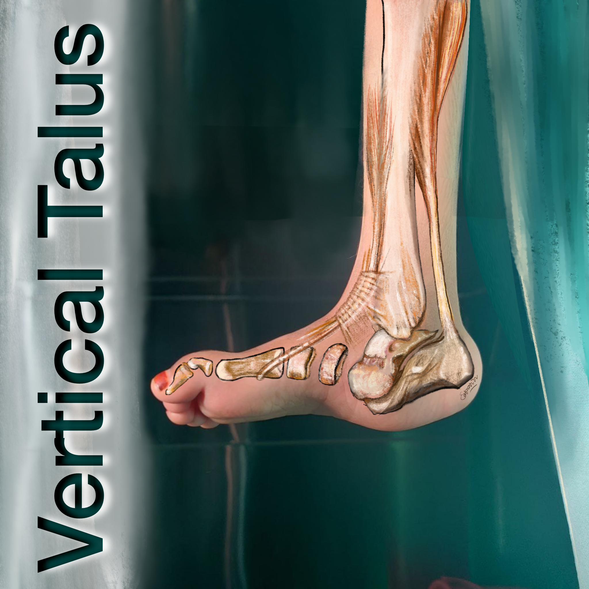
To help visualize this condition and bring attention to paediatric deformities, I created the artwork above as part of the ArztForACause project. This initiative uses visual art to raise awareness about orthopaedic conditions in children and helps fund essential care for those who need it most. Art has a unique way of making complex medical conditions easier to understand, bridging the gap between clinical diagnoses and human empathy.
What Exactly is Vertical Talus?
Congenital Vertical Talus (CVT) is a rare birth defect that affects the alignment of the bones in a baby’s foot.
In a typical foot, the talus (the ankle bone) points forward toward the toes, connecting the lower leg to the rest of the foot. In a child with CVT, the talus points downward toward the ground. This misalignment forces the bones in the middle of the foot to shift upward.
Because of this bone arrangement, the arch of the foot drops, and the sole becomes rounded outward. This creates a convex shape, which is why the condition is commonly called “rocker bottom foot,” as it mimics the curved bottom of a rocking chair.
Vertical talus is usually identified at birth or shortly after. The key physical signs include:
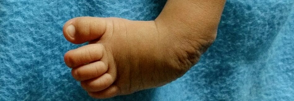
A Convex Sole: The bottom of the foot rounds outward instead of having an inward arch.
Upward Pointing Toes: The forefoot and toes point up and outward.
A Rigid Foot: Unlike some other newborn foot conditions which are flexible, a foot with vertical talus is stiff. You cannot easily gently bend the foot into a normal, flat position.
A Tight Achilles Tendon: The heel is pulled up tightly and does not touch the ground easily.
While the condition is not painful for a baby, it must be treated. If left uncorrected, a child will eventually walk on the inside ankle bone instead of the sole of their foot, leading to severe pain, calluses, and major difficulties with mobility and wearing standard shoes.
How is it Treated?
The goal of treatment is to realign the bones so your child can have a functional, pain-free, and stable foot.
In the past, treatment required extensive, invasive surgery. Today, the standard of care is much gentler and relies on minimally invasive techniques:
1. Serial Casting (The Dobbs Method): Treatment usually begins in the first few weeks of life. A paediatric orthopaedic surgeon will gently stretch the baby’s foot and apply a plaster cast. Every week, the cast is removed, the foot is stretched a little closer to the correct position, and a new cast is applied. This usually takes about 4 to 6 weeks.
2. Minimally Invasive Surgery: Once the casting has properly stretched the soft tissues, a minor surgical procedure is usually required. The surgeon will make a tiny incision to insert a small pin that holds the bones in the correct alignment. Often, a small release of the Achilles tendon (tenotomy) is also done to allow the heel to drop down.
3. Bracing: After the pin is removed (usually after a few weeks), the child will need to wear a special brace or special shoes connected by a bar. Bracing is crucial to ensure the foot does not shift back into the rocker-bottom shape as the child grows.
Moving Forward
A diagnosis of vertical talus is just a starting point. With a dedicated treatment plan and consistent bracing, the long-term outlook is excellent.
Dr. Easwar T. Ramani
Senior Consultant & Head of Paediatric Orthopaedics and Scoliosis
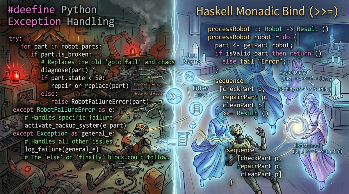
Imperative pyramid of doom versus the Monadic rescue.
PART 1 OF 6
Escaping the Imperative Pyramid of Doom
How a cerebral-palsy gait lab finally taught me what a Haskell Monad actually is
It started with one sentence. I had been teaching myself Haskell for weeks and making real progress — I had wrapped my head around currying and partial application, a milestone in its own right. Then, reading about the concept everyone warned was the final boss of the language – The Monad, I walked straight into this:
“A monad is just a monoid in the category of endofunctors, what’s the problem?”
— the sentence that stopped me cold
And I stopped. Not paused — stopped. Every word in it was a door into another locked room: monoid, category, endofunctor. Every tutorial I opened tried to pick those locks with even more abstract category theory, or with bizarre analogies about burritos. None of it clicked.
I didn’t need a burrito. I needed the Monad grounded in my reality: the messy, sweat-soaked, packet-dropping world of building a clinical gait-analysis pipeline for my cerebral-palsy clinic. I was building a software to analyse gait in children with cerebral palsy, my latest addition to the AuShadha Project. It was a 15 year dream, full of incremental learnings, stoppages, hurdles… I thought I had not hit another hurdle.
So I threw out the tutorials which were abstract, un-relatable and rebuilt the idea from the only place it ever makes sense to me — the lab floor.
This is Part 1 of a six-part journey. Over the series I’ll trace the Monad from a practical headache (this part), back to its 1950s mathematical origins (Part 2), down into its formal machinery (Parts 3 and 4), and finally through the infamous one-line definition that started this whole obsession (Part 5), before tying off the loose ends of Semigroups and Totality (Part 6).
We start where I started: drowning.
The premise: the messy reality of clinical data
Picture the setup.
A child with cerebral palsy walks on a gait lab path. I’m capturing markerless computer-vision joint angles at 120 frames per second and simultaneously ingesting high-frequency EMG from surface electrodes.
In a perfect, theoretical world, the data flows beautifully. I write a tidy function to calibrate a raw angle by adding a 5-degree offset:
But the clinic is not a perfect world. Wires come loose. Sensors drop packets. A parent leans in and occludes the camera. Sometimes I get a reading, and sometimes I get an error, or nothing at all.
Programmatically, this scenario is a nightmare. We have to prepare and programme so defensively, preparing for the edge cases, fear, hope and pray that some edge cases that we had not envisioned before would jump in, cause a runtime error and crash the programme. The traditional imperative programmer would check the scenarios that they can envision and prepare… and hope. This is very not very dependable.
This is the first thought process and fear that Haskell forced me to unlearn: we do not use `null` or None to represent missing data. Instead we use a context — a wrapper or a box or a container. The most common one is Maybe ( There are other containers in Haskell like Either, IO etc .. ). A value sits inside the container. A Maybe Float box can have two values inside it – Nothing or Just. A value of type Maybe Float is a box that is exactly one of two things:
Just 45.0 — the sensor worked; here is my wrapped reading.
Nothing — the sensor dropped the packet.
And immediately I had a problem.
I couldn’t feed Just 45.0 into calibrateAngle, because that function wants a raw Float, not a Float sealed inside a box. The Haskell compiler will yell at me. I needed a safe way to reach into the box, apply the calibration if the data existed, and put it back — and if the box was empty, just pass the emptiness along without crashing.
Step 1 — Discovering the Functor
That exact problem handed me my first tool: the Functor.
A Functor is simply any wrapper that lets you map a normal function over the data hidden inside it, using a function called fmap (or its operator <$>).
-- HASKELL

sensorReading :: Maybe Float
sensorReading = Just 45.0

calibratedReading = fmap calibrateAngle sensorReading

-- Result: Just 50.0
If the sensor had failed and handed me Nothing , fmap would have safely returned Nothing — no calibration attempted, no null-pointer exception, no crash.
More about fmap and it’s common poorer cousin – the map
It’s worth slowing down on fmap's type signature, because the whole idea is compressed into one line:
fmap :: Functor f => ( a → b ) → f a → f b
Read it aloud: give me a plain function `a → b`, and a value of type `a` wrapped inside some functor `f`, and I’ll hand back the result wrapped in that same `f`. If you’ve written Python or JavaScript, you already know one special case of this — the humble map over a list. The difference is reach. Ordinary map only knows how to walk a list; its type is pinned to one container:
-- HASKELL

map :: (a -> b) -> [a] -> [b] -- lists, and only lists
fmap :: Functor f => (a -> b) -> f a -> f b -- ANY functor
fmap is the same shape with the concrete [] generalized to any container that knows how to be mapped over — Maybe, Either, IO, a tree, even a parser. In fact, for lists the two are literally identical: fmap = map. So map isn’t a rival tool; it’s just fmap with the container hard-coded to a list.
The gait-lab payoff is concrete: the single operator <$> (the infix spelling of fmap) calibrates a reading whether it arrived as a Maybe Float from a flaky IMU, an Either String Float carrying an error reason, or an IO Float straight off the camera. I write the calibration once; fmap makes it run safely inside whichever box the reading happens to be trapped in.
Now, my instinct as a Python programmer screamed the obvious objection.
But why not just use if/else?
My immediate thought was: why can’t I just check the value with an if/else and be done with it? Why invent this whole Wrapper concept?
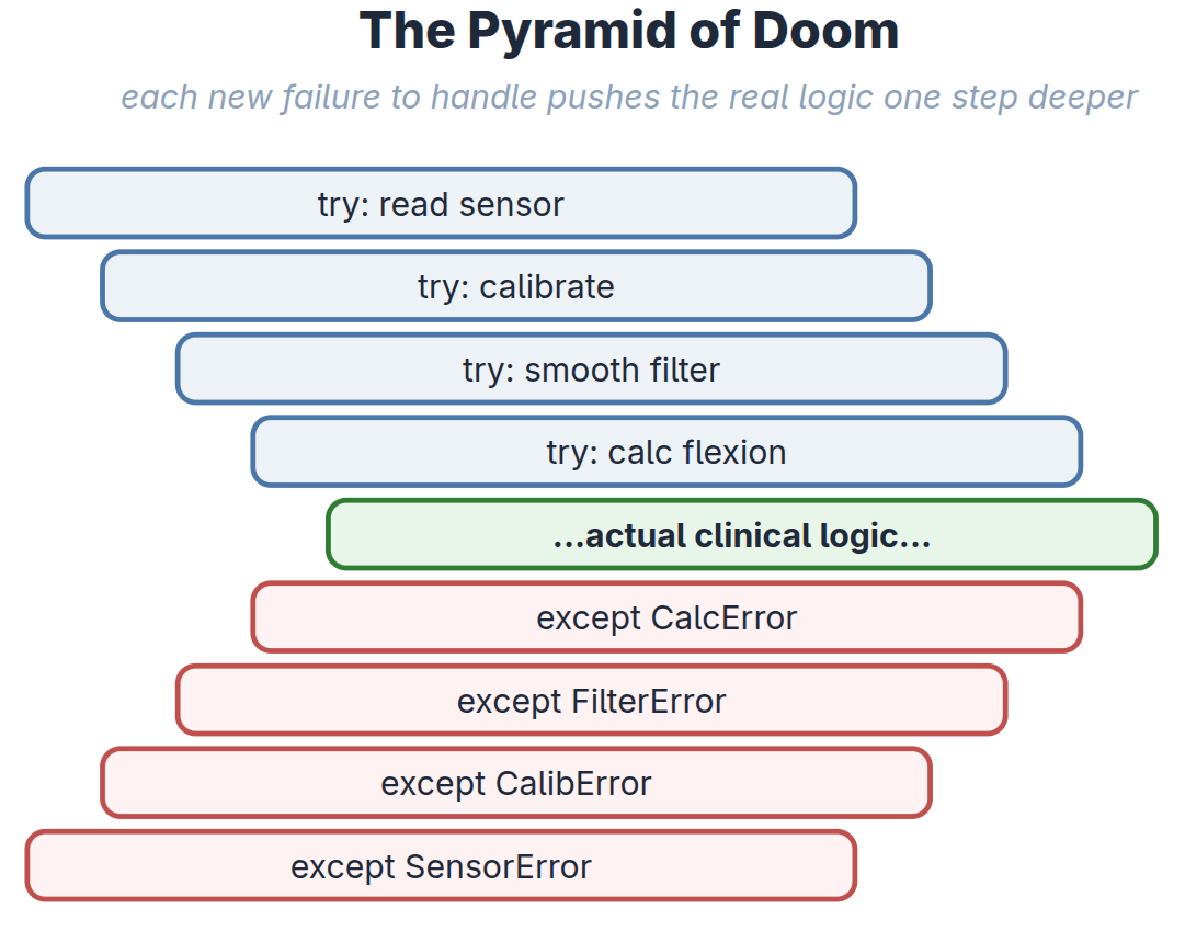
It comes down to scale and safety. If I use if/else for a multi-stage pipeline — read the sensor, calibrate, smooth, compute flexion — I end up with a nested pyramid that tangles clinical logic together with error-handling plumbing.
And here’s the point: the moment I try to be tidy and abstract that if/else into a reusable helper, I have accidentally reinvented the Functor.
fmap is not magic. It is just the standardized, universal version of the plumbing I was about to write by hand.
Step 2 — Applicatives and the two-sensor problem
In gait analysis I rarely care about one absolute angle; I care about the relative angle between two joints. So I need a function of two arguments — a thigh reading and a shank reading:
Both readings arrive messy, each in its own Maybe box.
When I tried fmap on just the first sensor, currying bit me: fmap applied the first argument and handed me back a function still waiting for the second argument — and it stuffed that half-applied function back inside a Maybe box. Now I had a wrapped function that needed to be applied to a wrapped value, and plain fmap couldn’t do it.
Enter the Applicative Functor and its operator <*>:
Just as the Functor abstracted the if/else for one wrapped value, the Applicative abstracts the nestedif/else for many wrapped values. If either sensor drops a packet, the whole chain short-circuits cleanly to Nothing .
A note on order of precedence in Haskell and why fmap does not work in multi-argument scenarios
finalAngle = calculateRelativeAngle <$> thighSensor <*> shankSensor Why are there no brackets — and what runs first?
That line looks ambiguous ( again that is me, coming from Python and expecting brackets to the right) . It isn’t. Both operators in it — <$> (which isfmap) and <*> — are declared infix : the same precedence level , and left-associative. Same precedence plus left-associative means Haskell groups the line strictly left-to-right, exactly as if I had reached for parentheses myself:
So the answer to the question on which runs first is : The functor step `<$>` runs first, on the left. It does not run the applicative first and then feed the result into calculateRelativeAngle.
Here is the actual two-step trace, taking thighSensor = Just 50.0 and shankSensor = Just 15.0:
Step 1 — `<$>` (fmap).calculateRelativeAngle takes two arguments, so applying it to only the first leaves a function waiting for the second. fmap puts that inside a box, producing a function trapped inside a Maybe:
HASKELL

calculateRelativeAngle <$> Just 50.0
==> Just (\shank -> 50.0 - shank) :: Maybe (Float -> Float)
That wrapped-up function is the dead-end that made fmap insufficient and forced us to reach for the Applicative in the first place.
Step 2 — `<*>` (Applicative) does the job fmap cannot: it applies a boxed function to a boxed value.
HASKELL

Just (\shank -> 50.0 - shank) <*> Just 15.0
==> Just (50.0 - 15.0) ==> Just 35.0
If either box were Nothing, the whole chain short-circuits to Nothing. Brackets aren’t required because the default left-to-right grouping is already precisely the one we want. (Function application binds tighter than any operator, so calculateRelativeAngle is a finished atom before <$> even looks at it.)
The people who chose these fixities did it deliberately: f <$> x <*> y <*> z is meant to read like an ordinary multi-argument call — left to right, no parentheses. That idiom has a name: applicative style.
Step 3 — The final boss: the Monad
I had handled wrapped data and multiple wrapped inputs. The final hurdle — the one that forced me to actually understand Monads — was this: what happens when my own function returns a wrapped value?
I wanted to validate that a computed angle wasn’t biomechanically impossible:
Please note that the function validateBiomechanics takes a Float and gives back a Maybe Float. So we cannot feed out previous data which is a Maybe container into this function as a direct argument. That is where Monadic operation comes in.
We need a way , an operator or tool, that can stand between our Maybe box and validateBiomechanics , take out the value out of the Maybe box , feed it to validateBiomechanics, accept another Maybe result from that, enclose it back in our original Maybe box out of which we took the value out and then since now we have two Maybe boxes, squish it down to just one Maybe box.
-- HASKELL

validateBiomechanics :: Float -> Maybe Float
validateBiomechanics angle =
  if angle >= -10.0 && angle <= 150.0
    then Just angle
    else Nothing
A confusion of Maybe, Nothing, and Just
The Type vs. Constructor Epiphany
For a while I was confused about Maybe, Just, and Nothing — functions promised a Maybe Float but returned a Just or Nothing. The line that unlocked it was the split between the type level and the value level: Maybe is the broad family name that exists only for the compiler; Just and Nothing are the actualshapes of data Maybe can hold. Because the compiler knows the Maybe family has exactly two members, it uses pattern matching to mathematically prove I’ve handled every possible outcome.
Can fmap do it ?
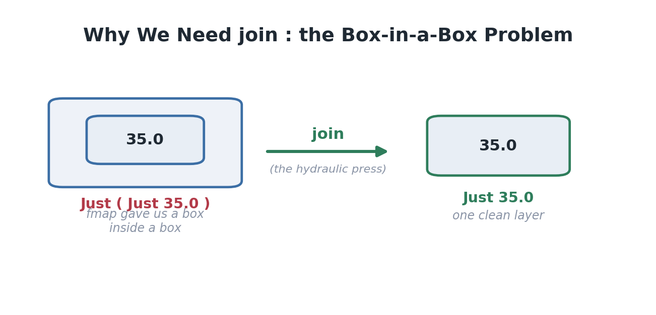
If I fmap this over Just 35.0 , fmap pulls out the 35.0 , feeds it to the function (which returns Just 35.0 ), and dutifully puts that result back in a box — leaving me with Just (Just 35.0) . Boxes inside boxes. I needed a tool that could take a wrapped value, feed it into a function that itself returns a wrapped value, and flatten the result so the boxes don’t stack up.
Please welcome our next tool, after fmap and applicative -- join
That flattening tool is the Monad bind operator , >>= :
-- HASKELL

cleanResult = Just 35.0 >>= validateBiomechanics
-- Result: Just 35.0   (one box, not two)
fmap alone leaves nested boxes; the Monad’s job is to flatten them back to one clean layer.
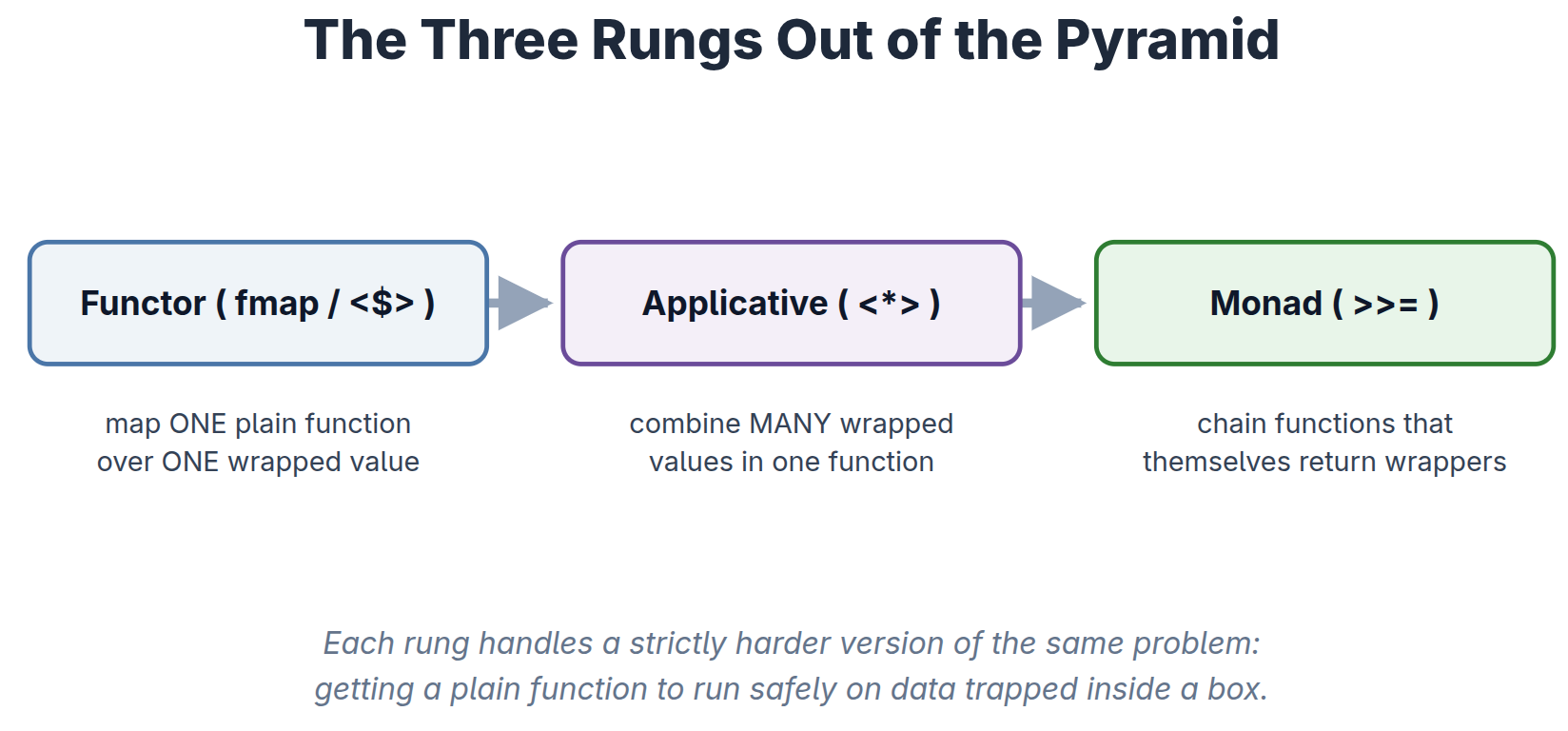
That’s the whole promise of a Monad, at the intuition level. Functor maps into a box. Applicative combines boxes. Monad chains functions that return boxes, flattening as it goes. Three rungs of the same ladder, each handling a strictly harder version of one problem.
The three rungs out of the pyramid. Each tool solves a harder version of “run a plain function on data trapped in a box.”
Unveiling the syntactic sugar: do notation
Chaining >>= operators by hand gets dense fast. This is exactly the tangled, rightward-marching mess I was trying to escape — the Pyramid of Doom that a Python programmer knows intimately:
Not convinced ?
Here is the how the full code may look like once functions are chained using >>=
Count the ways it fights you. Three lambdas march off to the right — a Pyramid of Doom rebuilt out of >>= — the clinical logic that actually matters ( thigh - shank ) is buried three indents deep in plumbing, and there’s a tail of ))) you have to balance by hand.
A hidden redundancy the math can spot
Look at the innermost step: validateBiomechanics relativeAngle >> (\validAngle -> return validAngle) . Feeding a value into >>= only to immediately return it does nothing — by the right-identity law (which we will talk about in Part 3) that whole line is exactly equal to :
validateBiomechanicsrelativeAngle on its own. The do block spells out the redundant final step for symmetry; the algebra says you could drop it. Monad laws aren’t decoration — they let you reason about and simplify actual pipeline code.
Every <- in the tidy version is one of these >>= (\x -> …) wrappers: the compiler generates precisely this nested-lambda tower and then mercifully hides it from me. (This is the literally what GHC performs on every do block, – more about this in Part 4)
So here is Haskell’s answer: do notation — syntactic sugar that unwinds that exact tower back into sequential-looking code, while the Monad handles all the failure branching invisibly underneath:
The `<-` operator is just >>= in disguise. It extracts the value from the box; if the box is empty, the whole block instantly aborts to Nothing.
`<-` versus `let`. I use <- to unpack monadic boxes, and let for plain, unwrapped math.
`return` is a trap for anyone coming from Python or C. It does not stop execution. It is just an ordinary function whose only job is to take a pure value and drop it into a default box. (We will speak about return again, maybe, in Part 3. Pun intended 😉 )
Step 4 — Making it real with Either and IO
Maybe can only say that something failed, never why. In a clinical setting, “it failed” is not an acceptable log line. So I upgraded to the `Either` Monad, which lets me carry the reason for failure alongside the failure itself:
-- HASKELL

validateVision :: Float -> Float -> Either String Float
validateVision angle confidence =
  if confidence > 0.85
    then Right angle
    else Left "Vision Error: Patient occluded."
Chained inside a do block, my pure math pipeline stays flawless: if the camera glitches, it returns a specific, human-readable error string instead of a crash. (This idea — a function that has a legal answer for every possible input, including failure — has a name, Totality, and it will be discussed probably in part 6)
But pure logic alone does nothing; a program with no side effects just warms up the CPU as many Haskellers proudly say. To actually touch the hardware and do something useful like announcing to the user on the monitor as an output and printing characters, I reach for the `IO` Monad, which quarantines the messy real-world boundary :
-- HASKELL

runGaitAnalysis :: IO ()
runGaitAnalysis = do
  putStrLn "Starting patient gait analysis..."
  rawAngle <- readCameraFeed            -- messy IO boundary
  visConf  <- readCameraFeedConfidence  -- messy IO boundary
  let result = calculateMuscleForce rawAngle visConf   -- pure math
  case result of
    Left err -> putStrLn ("FAILED: "  ++ err)
    Right f  -> putStrLn ("SUCCESS: " ++ show f)
And there it was. The Monad wasn’t a mystical entity or an internet punchline. It was the universal adapter that let me build one elegant, predictable pipeline — managing missing data, tracking clinical errors, and safely touching physical hardware — without a single line of spaghetti.
That’s the intuition. But intuition left me with a nagging question: where did this idea come from, and why did a purely functional language need rescuing by a piece of 1950s abstract algebra in the first place? In Part 2, I trace the Monad’s improbable journey from a mathematician’s chalkboard to my clinic’s compiler.
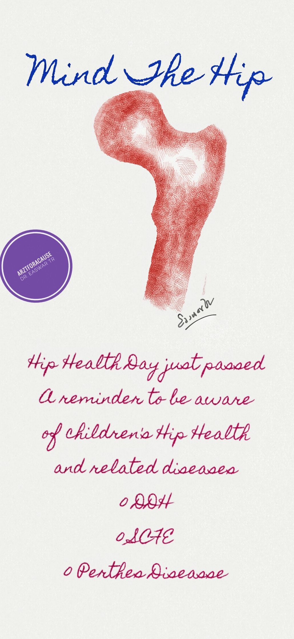
#HipHealthDay just passed on June04. A perfect time to remind ourselves if the importance of childs #hip #health and the common #diseases affecting childs hip joint.
Child hip health starts from the time a child is born. It is important to have your pediatrician examine you newborn to make sure the childs hip has no obvious signs of dislocation. This condition is called DDH and is often missed in newborn period. It’s easiest to treat early and is the cause of significant difficulty to the child later if undetected.
In the newborn period it’s very easy and reassuring to do an ultrasound screening of the hips to rule out a hip dislocation or a more subtle variation called ‘dysplasia’ which is not possible to pick up via examination.
In older children hip disease usually has pain or limp. Be very suspicious if your child has a limp without a fall to account for it. Don’t ignore pain around the hip, thigh or knee. It is important to be aware that the child may have pain in the knee or thigh instead of the hip in hip joint problems
If the child is walking with a limp, if you feel that the child has a difference between the limb lengths or swaying from side to side it may be an indication of a hip problem.
Don’t hesitate to consult your #pediatric #orthopedic #surgeon
So , Mind the #Hip !
– Dr. Easwar Tr . Paediatric Orthopedic Surgeon Baby Memorial Hospital , Kozhikode Kerala
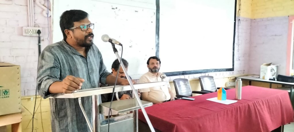
It was so nice to meet and talk to the parents about #cerebralpalsy and #developmentaldelay in #children at the #puthur #brc in #palakkad yesterday
Stress was on the need for #earlyintervention and adherence to #treatment especially #physiotherapy. The challenges parents face while continuing care is an eyeopener everytime I hear them out.
As in most diseases, treatment challenges in #cerebralpalsy is more to do with #social, #familial and #accessibility issues than actual lack of desire.
Most parents are willing to face the #financial challenges if it means the child will improve. Many are disillusioned with existing treatment methods and fall out because of slow progress, lack of motivation from professionals around and also pure financial pressure. Many have siblings who are healthy and would like to divert their limited resources to that child. This is such an unfortunate choice , but many parents make it.
Such talks and interaction are a wonderful opportunity to break the ice, motivate and also clear their apprehensions on the treatment of cerebral palsy.
A big thanks to Puthur BRC for organising this meeting. More to follow 🙂
Attaching below a few pictures and also a short video which the BRC officials had shared.
Mr. Shivakumar BPC, Puthur BRC Parents were enthusiastic and participated wellA compilation of photos of the day
Before the pandemic struck and wrought drastic stop to treatment of children with cerebral palsy.
Two years have passed and we hobble back to restart our work and pick up pieces. Many children have lost the improvements that they had attained as they lost out of treatment due to lack of money, travel restrictions and physiotherapy.
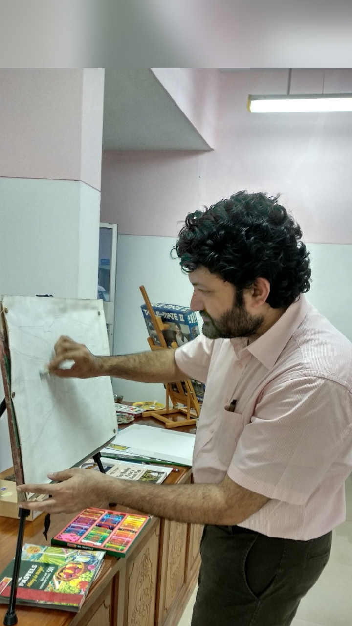
This is October 2019 we hosted a live painting workshop at ICCONS to raise awareness about Cerebral Palsy.





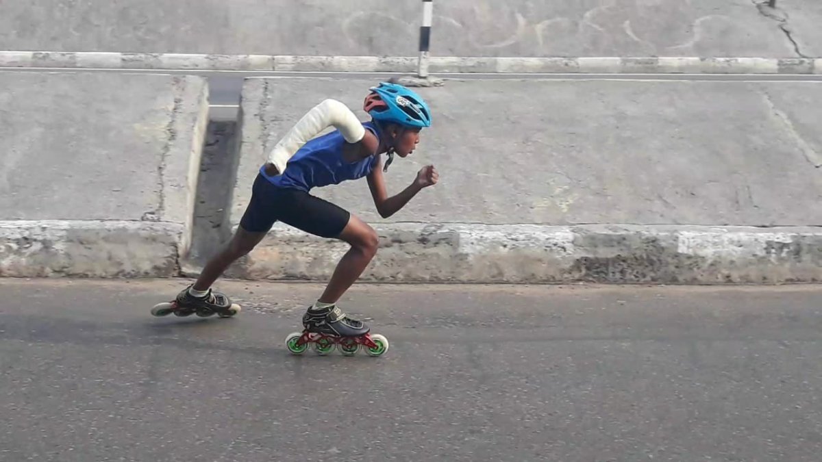
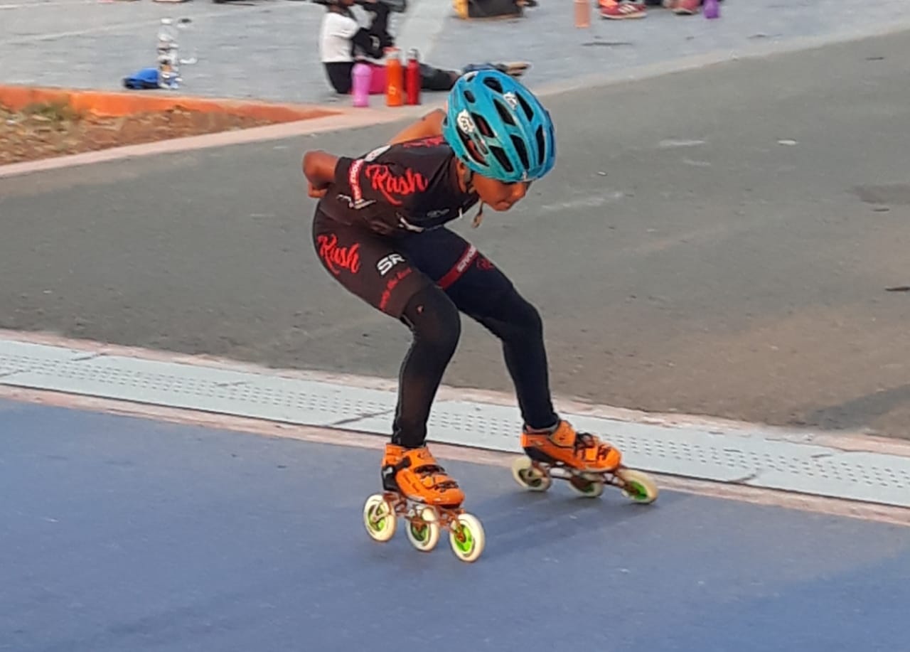
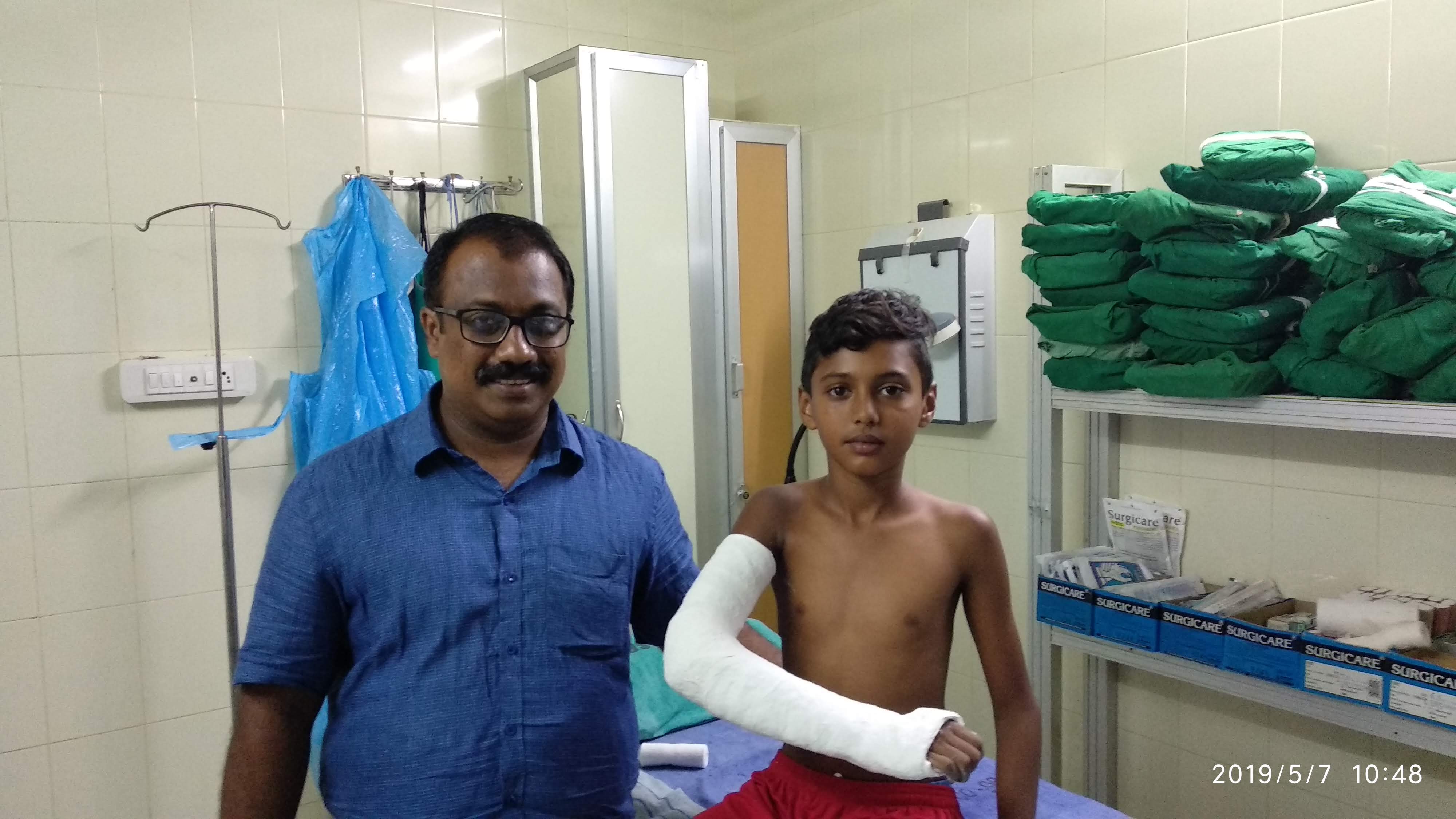 Aniruddhan with his father. His latest fracture a minor blow in his quest
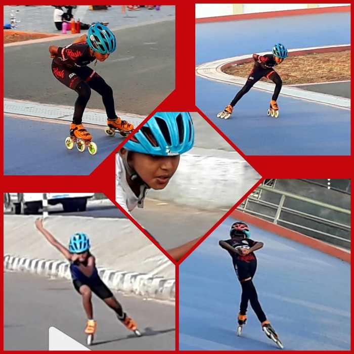 Aniruddhan V , on Roller Skates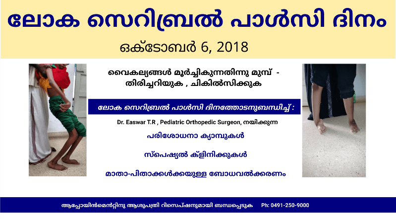

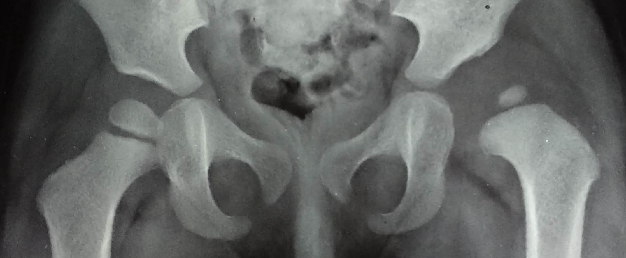









 Dr. Easwar T.R